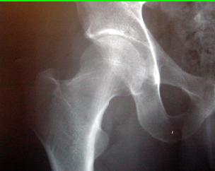
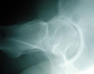
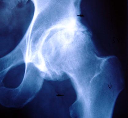
Osteoarthritis is a condition that affects a significant proportion of the population. Essentially, it is a condition in which the cartilage that ‘covers’ the bones (articular cartilage) degenerates. For this reason, osteoarthritis is commonly referred to as ‘wear and tear arthritis’.
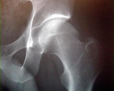
The articular cartilage is a particularly specialised structure, giving joints a fantastic smooth and friction-free movement. As the cartilage degenerates bone can become exposed on both sides of the hip joint. This can result in pain. The condition can cause swelling within the joint (an effusion) that can result in stiffness and discomfort. As the condition progresses, spurs of bone (osteophytes) can form around the rim of the joint. This process can further restrict movement and can cause pain as one part of the bone impinges on another during ‘normal’ movement.
If the condition is allowed to progress further, the underlying bone can start to become damaged. Typically, this causes worsening pain, more restricted movement and can result in deformity in the alignment of the hip joint. Deformity can also occur secondary to soft tissue contractures. In the hip, a combination of shortening, soft tissue contracture, pain and indeed weakness can result in a limp.
The overall effect of this breakdown of the articular cartilage in the joint is pain, stiffness, restricted mobility and frequently a limp. All this can affect how a patient is able to go about their normal, everyday domestic and occupational activities as well as their ability to enjoy recreational hobbies or sports.
The progress of the changes in osteoarthritis, described above, can be seen in the series of x-rays below: