The hip is a large, versatile and stable joint allowing an excellent range of movement with very low concerns with instability. Surrounded by the large muscle groups that initiate movement and generate power, hip function is also contributed to by ligament attachments and by the pelvic musculature. The hip transmits load from the axial spine via the pelvis into the legs. As such, the hip is extremely important in all activities of daily life. Huge loads are transmitted through the hips as we perform our required and desired activities. These can be up to seven times your body weight during activities such as running up stairs.

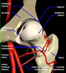
As can be seen from the diagram, the hip comprises an acetabular socket within the pelvis. The bone in the socket is covered with a lining of a highly-specialised and durable layer called the ‘articular cartilage’. The socket itself is subsequently ‘deepened’ by a rim of a more fibrous cartilage, the labrum, that is circumferentially attached to the bony rim, further enhancing stability.
On the other side of the joint is the femoral head. The femoral head functions as a ball on top of the proximal femur. Again, the bony surface is covered with a lining of the same ‘articular cartilage’ to give an extremely smooth bearing surface against the articular cartilage of the acetabulum. The hip joint is lubricated by fluid produced by the synovial soft tissues that line the capsule of the joint. This synovial fluid is produced and resorbed in perfect balance. The fluid both lubricates the joint and maintains articular cartilage integrity.

A combination of the bony anatomy, specialised articular cartilage linings, lubricating fluid enclosed within the soft tissue capsule and moved by the surrounding muscle groups, allows a wide range of pain-free movement with excellent control and significant power in the non-diseased hip.
The capsule extends some way down the neck of the femur as is demonstrated in the diagram. This capsule surrounds the joint and contains the joint fluid. The hip capsule itself has stretch sensors within it. It is these sensors that detect an increase in joint fluid, ‘swelling’ within the capsule, as can occur in the early stages of arthritis or following injury. This ‘stretch’ of the capsule can result in a sensation of stiffness and discomfort in the hip without any obvious external signs as the hip joint itself is located deep beneath the muscles that move it.
There are a number of muscle groups that cross the hip joint, attaching both to the bony pelvis and to the proximal femur. These muscle groups allow us to move the hip with wonderfully-detailed control and power to facilitate the excellent range of movements that are possible in the hip.
The main muscles that flex the hip are the psoas, the anterior part of the gluteus medius muscle and the quadriceps muscles that attach to the anterior part of the proximal femur.
There are a group of muscles on the inside of the thigh, the ‘adductors’, that move the hip in towards the mid-line. Similarly, there are very important muscles that extend from the lateral part of the pelvis to the tip of the greater trochanter of the femur. These ‘abductor’ muscles are extremely important. They allow us to walk with our pelvis level, even when all our weight is actually on one leg. It is frequently weakness in these hip abductors, as a consequence of pain or following surgery, that can result in a limp. Rehabilitation of the abductor muscles is important following hip surgery.
Posteriorly, the muscles that are responsible for extending the hip, such as the gluteus maximus and the hamstrings, attach onto the posterior aspect of the pelvis and the posterior proximal femur.
All of these various muscle groups are essential in maintaining normal hip function.
Frequently, as a consequence of discomfort resulting in decreased activity, these muscle groups are weak and can be wasted. It is for this reason that physiotherapy can be beneficial to patients with early arthritis, maintaining muscle strength and support as well as restoring mobility. This treatment can also optimise and support the articular cartilage lining of the hip joint.
Similarly, all these muscle groups need to be ‘worked on’ following surgery once the painful and/or stiff hip joint has been treated. Rehabilitation with exercise and physiotherapy reconstitutes the muscle bulk, improving strength and helping to achieve an optimal and functional recovery.